UPPER GASTROINTESTINAL HAEMORRHAGE
| Key point |
In 7% of cases the procedure was performed too late to benefit the patient.
Please note that this value was originally published incorrectly as 79% |
To identify patients who had suffered from upper GI haemorrhage, NCEPOD identified patients who had laser destruction or cauterisation of their lesion, or where ‘other’ procedure suggested treatment for haemorrhage. From this 65% (524/809) of patients were identified as having suffered an upper (GI) haemorrhage.
Urgency of the procedure and patient’s physical status
(back to top)
In 1995 Rockall and co-workers1 devised a scoring system for the risk of rebleeding and death after acute gastrointestinal bleeding, which is widely used by upper GI specialists in guiding their local protocols. The risk factors that accurately predict death at the time of admission are summarised here (from the BSG in their guidelines for the management of non-variceal upper GI haemorrhage)3 :
• |
Age: death in patients less than 40 years is rare while the risk of death is 30% in those over 90 years of age |
• |
Comorbidity: particularly advanced renal or liver disease, or disseminated cancer. But it is crucial that diseases of the heart, respiratory system and central nervous system are recognised and appropriately managed |
• |
Shock: defined as a heart rate >100 beats/min and systolic blood pressure <100mmHg |
• |
Endoscopy findings: Mallory Weiss tear or finding no stigmata of recent haemorrhage are low risk whereas active bleeding in a shocked patient carries a 80% risk of continued bleeding or death. |
For those with liver disease the prognosis is related to the severity of liver disease rather than to the magnitude of haemorrhage.
With these factors in mind NCEPOD analysed the urgency of the procedure (Table 51)
and the patient’s physical status (Figure 17).
| Table 51. Urgency of the procedure for upper GI haemorrhage
|
| Urgency |
|
Elective/scheduled |
|
| Urgent |
|
| Emergency |
|
| Sub-total |
|
| Not answered |
|
Total |
|
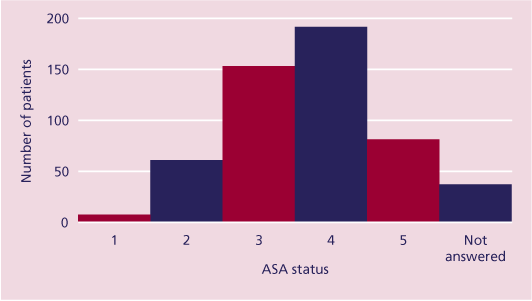
Figure 17. ASA status of those with upper GI haemorrhage
Of the 524 upper GI endoscopies for haemorrhage, 50% (243/488) were classed as urgent and 36% (175/488) as emergency (Table 51). Most of the patients were also assessed as being either severely ill (ASA 3), 31% (154/492), having a severe illness that is a constant threat to life (ASA 4), 38% (188/492), or moribund (ASA 5 ), 16% (80/492). Four patients who were classified as ASA 5 were apparently having a scheduled procedure. Table 52 presents the comorbidities at the time of the procedure.
| Table 52. Comorbidities for upper GI haemorrhage (answers may be multiple)
|
| System |
Total
n = 515 |
| Respiratory |
148 |
| Cardiac |
173 |
| Neurological |
128 |
| Hepatic |
59 |
| Renal |
122 |
| Total |
630 |
| None |
18 |
| Not answered |
9 |
Of these, 24% (123/515) had ischaemic heart disease and 15% (77/515) had an acute
chest infection. Cardiac disease in association with an upper GI haemorrhage should
indicate the need for ECG monitoring, and respiratory disease the need for pulse oximetry and supplemental oxygen during the endoscopy (see earlier chapter entitled ‘Sedation
and monitoring’). From a separate question 38% (199/524) of patients with upper GI haemorrhage had cirrhosis. The Childs-Pugh score for those patients with cirrhosis is presented in Table 53.
| Table 53. Childs-Pugh score of those patients with cirrhosis and upper GI haemorrhage
|
| Childs-Pugh Score |
|
A |
|
| B |
|
| C |
|
| Sub-total |
|
| Not answered |
|
Total |
|
The high proportion of patients with advanced cirrhosis reinforces the association between the severity of liver disease and death following upper GI haemorrhage. Table 54 presents the anticipated risk of death in the opinion of the clinician at the time of the procedure.
| Table 54. Anticipated risk of death for upper GI haemorrhage
|
| Risk of death |
|
Not expected |
|
| Small but significant risk |
|
| Definite risk |
|
| Expected |
|
| Sub-total |
|
| Not answered |
|
Total |
|
In total, 89% (454/508) had a definite risk of death or death was expected indicating that the sample contained an exceptionally high-risk group of patients.
Appropriateness of the procedure and organisation of care
(back to top)
In their review of the cases NCEPOD advisors assessed the appropriateness and timing of the procedure for those with upper GI haemorrhage. Table 55 presents the advisors' opinion on the appropriateness of the procedure and Table 56 the reasons why the procedure was considered inappropriate.
| Table 55. The appropriateness of the procedure for those with upper GI haemorrhage
|
| Procedure appropriate |
|
Yes |
|
| No |
|
| Undecided |
|
| Sub-total |
|
| Insufficient information |
|
| Not answered |
|
Total |
|
| Table 56. Reasons for inappropriate procedures (answers may be multiple)
|
| Reason |
Total
n = 23 |
Futile procedure |
13 |
| No endoscopic procedure indicated |
4 |
| Different endoscopic procedure indicated |
2 |
| Surgery in the first instance would have been more appropriate |
1 |
| Other |
6 |
| Total |
26 |
| Not answered |
2 |
It is reassuring that for 92% (473/515) of cases the procedure was assessed as appropriate. For these 473 cases the advisors were asked to consider whether the procedure was also appropriately timed. In 33/473 (7%) of cases the timing was considered inappropriate and the reasons for this are presented in Table 57.
| Table 57. Reasons why the timing of the procedure was inappropriate (answers may be multiple)
|
| Reasons |
Total
n = 31 |
Late, delayed referral |
10 |
| Late, inappropriate prolonged resuscitation |
5 |
| Late – other |
16 |
| Early, further preoperative resuscitation indicated |
4 |
| Early – other |
2 |
| Total |
37 |
| Not answered |
2 |
On reviewing the cases NCEPOD advisors were also concerned about delays for a variety
of non-clinical reasons, for example there were 21 delays for organisational issues, including nine cases that should have been done as emergencies but were deferred until normal working hours. There were also 20 cases where there may not have been delays, but there were other concerns about the organisation of care. Table 58 presents the advisors' opinion on the overall quality of care.
| Table 58. Advisor opinion on the overall quality of care provided for upper GI haemorrhage
|
| |
|
Good practice |
|
| Room for improvement |
|
| Less than satisfactory |
|
| Sub-total |
|
| Insufficient information submitted to assess |
|
| Not answered |
|
Total |
|
The reasons for less than satisfactory care were diverse. They were clinical, e.g. inadequate or delayed resuscitation, or organisational, e.g. lack of ICU beds, poor referral policies, inadequate out-of-hours care etc. However, that there was room for improvement or the care provided was less than satisfactory in 27% of patients who suffered upper GI haemorrhage suggests that Trusts should review the provision of care for these patients in order to identify deficiencies locally.
| Case Study |
|
| An elderly patient with cirrhosis (no cause stated) and ischaemia related biventricular failure presented with a haematemesis that was not considered to be severe by the admitting clinican as the “urea is only 6.5”. The patient was tachypnoeic, tachycardic and hypotensive. Before endoscopy, the patient did not receive either supplemental oxygen or intravenous fluids – which in view of the cardiac condition should have been governed by central venous monitoring. |
The clinician failed to consider the:
1. effect of liver disease on blood urea levels
2. significance of a tachycardia and hypotension
3. likelihood of abnormal clotting, and no test was requested until two days after admission.
| Case Study |
|
| A young patient with alcoholic cirrhosis presented after a haematemesis and melaena. On examination the patient was confused, tachycardic and hypotensive. Initial results included a glucose of 2.6 mmol/l, Hb 5gm/dl, platelets 30x109/L and an INR of 2.6. The pre-endoscopy treatment was resuscitation with normal saline, gelofusin and blood, and intravenous glypressin. Unfortunately no supplemental oxygen was given, and the hypoglycaemia, throbocytopenia and prolonged INR were not corrected. |
Specialty and grade of endoscopist (back to top)
| Key point |
The endoscopists managing patients with upper GI haemorrhage were mostly
of an appropriate specialty. However, 24% were trainees. |
The BSG guidelines recommend that patients admitted with upper GI bleeding should be the responsibility of a medical or surgical gastroenterologist who collaborates with a consultant in the other discipline. Ideally, specialist gastroenterologists (physicians or surgeons) should admit and manage these patients3.
| Table 59. Specialty of senior endoscopist for upper GI haemorrhage
|
| Specialty |
|
Specialised GI physician |
|
| Other physician |
|
| Specialised GI surgeon |
|
| Other surgeon |
|
| Radiologist |
|
| General practitioner |
|
| Nurse practitioner |
|
| Other |
|
| Sub-total |
|
| Not answered |
|
Total |
|
As can be seen from Table 59, 94% (482/512) of patients with upper GI haemorrhage had their endoscopy performed by a specialised GI physician (80%) or surgeon (14%).
| Table 60. Grade of senior endoscopist for upper GI haemorrhage
|
| Grade |
|
Consultant |
|
| Staff grade and associate specialist |
|
| General practitioner |
|
| Nurse practitioner |
|
| SpR - year 3 or over |
|
| SpR - year 1/2 |
|
| SHO |
|
| Other trainee |
|
| Sub-total |
|
| Not answered |
|
Total |
|
Consultants did most of the endoscopies (68%, 351/517) (Table 60). Most, but not all, of the remainder were done by specialist registrars (23%, 121/517), and staff grade and associate specialists (7%, 37/517). That one fifth of these very sick patients were done by SpR year 3 or over looks to be too high, but that would also depend on the time of the procedure and the level of supervision. The SpRs year 1/2 and the SHO were all physicians. Although their experience is not known one would suspect that emergency GI endoscopy for haemorrhage carried out by these grades would be inappropriate.
Location, sedation and monitoring (back to top)
| Key points |
13% of patients with upper GI haemorrhage received excessive sedation.
23% of patients received insufficient monitoring during the procedure. |
Most patients presenting with upper GI haemorrhage (87%, 448/516) had their endoscopy
in an appropriate location (Table 61) according to BSG recommendations3. However, it is also likely that the critical care areas (ICU/HDU) have facilities similar to those in a theatre environment. Thus, the location would seem appropriate in 98% of cases (506/516).
This figure may be even higher, but there are no specific details about facilities in the
remaining locations.
| Table 61. Endoscopy location for those with upper GI haemorrhage
|
| Location |
|
Dedicated endoscopy suite |
|
| Operating theatres |
|
| ICU/HDU |
|
| Admissions ward |
|
| A&E |
|
| Day surgery unit |
|
| X-ray department |
|
| Other ward |
|
| Sub-total |
|
| Not answered |
|
Total |
|
Endoscopists differ in their use of analgesia and sedation, based on personal preference, experience and the clinical condition of the patient. In the context of upper GI haemorrhage the combinations used are shown in Table 62.
| Table 62. Analgesia and sedation during endoscopy for those with upper GI haemorrhage (answers may be multiple)
|
| Analgesia and sedation |
Total
n = 477 |
| Local anaesthesia |
167 |
| Intravenous benzodiazepine |
308 |
| Intravenous opiod |
46 |
| Other intravenous sedation |
36 |
| Total |
557 |
| None |
28 |
| Not answered |
47 |
In interpreting these data it should be remembered that the endoscopist did not sedate all patients. For example those on ICU/HDU may have been in receipt of sedation and IPPV and those in the operating theatres may have had an anaesthetist present. This may account for some cases where no sedation was given and that the other intravenous sedatives included propofol, which is usually given by an anaesthetist. It is of concern that almost one third of these patients who by the evidence of their physical status and anticipated risk were very sick, received local anaesthesia to the oropharynx, including 25% (20/80) of patients who had an ASA status of 5. Local anaesthetic to the oropharynx alone may be appropriate for a sick patient. 30% (167/557) had local anaesthesia alone but 25% (139/557) had local anaesthesia combined with sedation. The risk of local anaesthesia to the oropharynx and aspiration is discussed further in the chapter entitled Sedation and monitoring'.
In 13% (68/524) of cases the advisors thought the sedation provided was inappropriate, mostly because of excessive benzodiazepine. 9% (49/524) of patients required reversal of their sedation with flumazenil and/or naloxone following the procedure, mostly because of sedation overdose, and this figure is too high. In 23% (123/524) of cases the advisors thought that there were deficiencies in patient monitoring. The reason for deficiencies is presented in Table 63.
| Table 63. Reasons for deficiencies in monitoring for those with upper GI haemorrhage (answers may be multiple)
|
| Reasons for deficiencies in monitoring |
Total
n = 123 |
No pulse oximetry |
6 |
| No ECG recording |
103 |
| No BP recording |
65 |
| No dedicated person to monitor patient |
5 |
| Other |
3 |
| Total |
182 |
| Not answered |
2 |
As discussed more fully in the chapter on ‘Sedation and monitoring’ the under use of ECG and blood pressure monitoring in these patients, who have the potential for haemodynamic instability, represents poor monitoring practice. 4% (19/495) of patients were not given supplemental oxygen during the procedure and this is clearly unacceptable.
The location of patients immediately following the procedure is presented in Table 64.
| Table 64. Location of patient immediately after the procedure for those with upper GI haemorrhage
|
| Location |
|
Dedicated recovery area within the endoscopy unit |
|
| Dedicated recovery area within the operating theatres |
|
| ICU/HDU |
|
| General ward |
|
| Died during the procedure |
|
| Other |
|
| Sub-total |
|
| Not answered |
|
Total |
|
It is unacceptable that any patient who has had an endoscopy for upper GI bleeding, particularly if they have received sedation, should go to an area without full recovery and resuscitation facilities such as a general ward.
Death and audit (back to top)
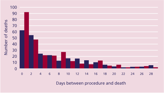
Figure 18. Days between procedure and death in cases with upper GI haemorrhage
Deaths within 72 hours (41%, 209/514) of the endoscopy (Figure 18) were likely to be due to either continuing haemorrhage, or associated complications. Deaths in the second week were probably mainly due to sepsis and organ dysfunction, and organ dysfunction was likely to be the main cause in the remainder of the thirty days.
Table 65 presents whether the department of the endoscopist who undertook the procedure held audit/morbidity/mortality meetings and Table 66 presents whether the case was considered at a meeting.
| Table 65. Were audit meetings held in the department of the endoscopist?
|
| |
|
Yes |
|
| No |
|
| Sub-total |
|
| Not answered |
|
Total |
|
| Table 66. Were deaths considered at an audit meeting?
|
| |
|
Yes |
|
| No |
|
| Sub-total |
|
| Not answered |
|
Total |
|
Of the 305 cases not considered at an audit/morbidity/mortality meeting it was intended to discuss 62 at a later date. However, this leaves 59% of cases where a patient died following an upper GI haemorrhage not being discussed at audit and a further 22% where audit was not specified.
|