| |||||
| |||||
|
PATIENT PROFILE
The age of all patients who underwent OGD is presented in Figure 16. 61% (493/809) were aged 70 years or older and 60% (485/809) were male.
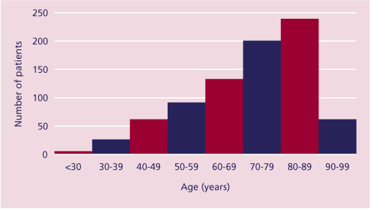
Figure 16. Age profile of patients undergoing OGD procedure
The diagnoses providing the reason for OGD, which were analysed from assessment of the free text for the procedure performed, are presented in Table 48.
| Table 48. The reason for OGD | ||||||
| Diagnosis |
|
|||||
Variceal disease |
|
|||||
Ulcer |
|
|||||
|
|
|||||
Benign
|
|
|||||
Polyp |
|
|||||
| Sub-total |
|
|||||
| Not answered |
|
|||||
Total |
|
|||||
The commonest diagnosis, 44% (355/807), was of variceal disease and in only 20% (163/807) was there a diagnosis of ulcer. This was surprising as ulcer disease is very common, and bleeding ulcers are more common than varices1. However, it suggests that patients with bleeding ulcers are more likely to survive compared with those who suffer variceal haemorrhage. This may reflect the severity of shock associated with variceal haemorrhage and the associated comorbidity, particularly hepatic disease1.
The complications or events that occurred within 30 days of the procedure are presented in Table 49.
| Table 49. Complications after therapeutic upper GI endoscopy (answers may be multiple) | |
| Complication | Total |
| Progress of medical condition | 203 |
| Respiratory problems | 175 |
Haemorrhage |
141 |
| Cardiac problems | 111 |
| Renal failure | 81 |
| Hepatic failure | 67 |
| Sepsis | 45 |
| Subsequent related surgery | 30 |
| Haematological problems | 30 |
| Electrolyte imbalance | 25 |
| Viscus perforation | 21 |
| Stroke | 14 |
| Total | 850 |
| None | 93 |
| Not answered | 69 |
This shows that, other than the progress of the medical condition, the most common complications were respiratory problems 21% (175/850), haemorrhage 17% (141/850) and cardiac problems 13% (111/850). These data are similar to previous studies that have shown cardio-respiratory complications to be the most prominent4 5 following both therapeutic and diagnostic upper GI endoscopy. The complications of viscus perforation and haemorrhage and the need for surgery may be related to the procedure, and these are presented in
Table 50.
| Table 50. Procedure versus operative complication (answers may be multiple) | ||||
| Procedure (total performed) | Perforation |
Haemorrhage |
Surgery |
Total n = 809 |
| Snare (5) | 1 |
0 |
1 |
2 |
| Coagulation (104) | 0 |
23 |
6 |
29 |
Laser (9) |
0 |
1 |
0 |
1 |
| Sclerosis (400) | 1 |
71 |
15 |
87 |
| Banding (41) | 0 |
6 |
0 |
6 |
| Dilation (125) | 15 |
5 |
0 |
20 |
| Stenting (259) | 4 |
35 |
8 |
47 |
| Total | 21 |
141 |
30 |
192 |
The techniques used to secure haemostasis were argon/plasma coagulation, laser treatment, injection with either adrenaline or sclerosing agents, or banding, or a combination of these techniques. Most were used appropriately and followed BSG guidelines2 3. However, in a number of patients with bleeding oesophageal varices adrenaline was injected into either the oesophageal mucosa and /or the adjacent varix in an attempt to control bleeding, which is not recommended in the UK guidelines of management of variceal haemorrhage2.
Although continued or recurrent haemorrhage can occur after haemostatic attempts with either argon/plasma coagulation (22%, 23/104), or sclerotherapy (18%, 71/400), or banding (15%, 6/41), there appeared to be a surprisingly high incidence following stenting (14%, 35/259).
The following case illustrates that repeat endoscopy can be indicated, and the fact that different pathologies can co-exist.
| Case Study | |
| A young patient with known cirrhosis presented with haematemesis. The pre-endoscopy management was exemplary. Subsequent endosopy revealed bleeding varices, which were banded. The patient received terlipressin, but continued to bleed. A further endoscopy 12 hours later confirmed that the treated varices were not bleeding, but there was a haemorrhage from a duodenal ulcer. Despite surgical intervention the patient continued to bleed, and died from disseminated intravascular coagulation. | |
| (back to top) |